← All services  /  SERVICE / MEDICAL AI

Governed medical AI for clinical workflow.

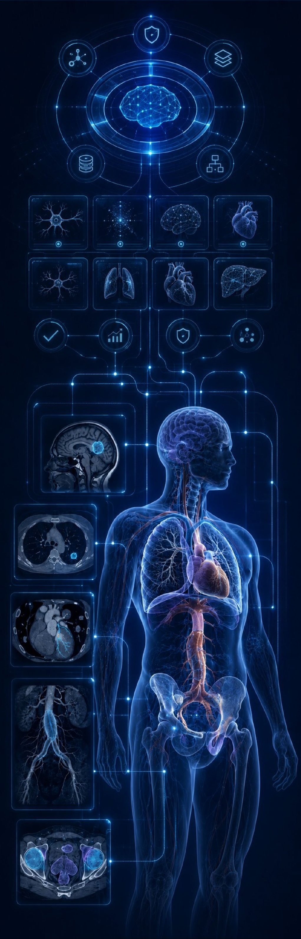
One governed platform and a focused portfolio of clinical AI, from certified brain-tumor detection to cardiovascular, thoracic and radiotherapy imaging.

3 approved TFDA devices
1 FDA 510(k) clearance
6 next products in certification
4 clinical domains

Technology partner: Shangya Technology Co., Ltd., Taipei, Taiwan

Medical AI across brain, chest, cardiovascular and pelvic imaging

THE PLATFORM

Liger turns medical AI into a managed clinical service.

Liger MedAI Management Platform connects imaging, AI orchestration, results and governance in one operating layer. TFDA Class II · No. 008624

  1. 01

    DICOM study

    Imaging studies leave the modality and PACS.

  2. 02

    AI orchestration

    Each study is routed to the right AI service.

  3. 03

    Physician review

    Every output stays under clinician review.

  4. 04

    Results to PACS

    Findings return to the normal reading workflow.

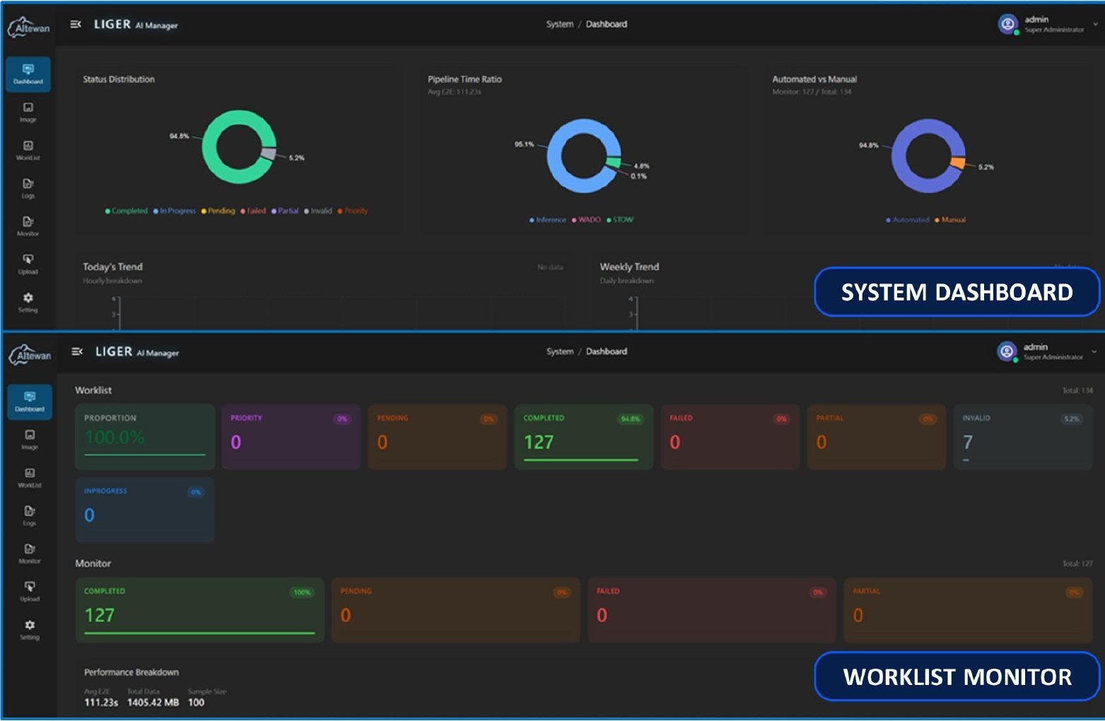
Liger AI Manager system dashboard and worklist monitor
Liger AI Manager: system dashboard and worklist monitor.

Clinical integration

PACS / DICOM connectivity routes studies to the appropriate AI service and returns results into clinical workflow.

Operational control

Multi-vendor orchestration, role-based access, reporting and audit functions support repeatable hospital deployment.

Interoperability

DICOM and FHIR for clinical-data exchange. One platform, multiple AI modules.

LIFECYCLE GOVERNANCE

Governance continues after deployment.

A closed loop from controlled introduction to monitoring, improvement and safe retirement, keeping performance trends, model stability and abnormal alerts visible without displacing physician review.

  1. 01

    Apply

    Create a controlled entry point for pilot or production use.

  2. 02

    Review

    Assess ethics, clinical benefit, risk and evidence.

  3. 03

    Deploy

    Define SOPs, access, integration and site rollout.

  4. 04

    Monitor

    Track performance, stability and operational alerts.

  5. 05

    Improve

    Investigate anomalies and govern validated updates.

  6. 06

    Retire

    Preserve evidence and a recoverable end-of-life path.

PORTFOLIO

Eight products across four clinical domains.

Approved products and products in certification are identified with the same evidence-led status system.

Brain MRI

  • DeepBT® Detector Approved · TFDA 007906
  • DeepBT® Detector-Plus Approved · FDA K252190 · TFDA 008460
  • DeepBT® Detector A+ TFDA review
  • CereMap TFDA preparation

Cardiovascular CT

  • DeepLDCT-CAC TFDA preparation
  • DeepAorA Detector TFDA review

Thoracic CT

  • DeepPulmo TFDA preparation

Radiotherapy CT

  • DeepPelvis TFDA preparation

CERTIFIED CORE PRODUCT

DeepBT® Detector-Plus

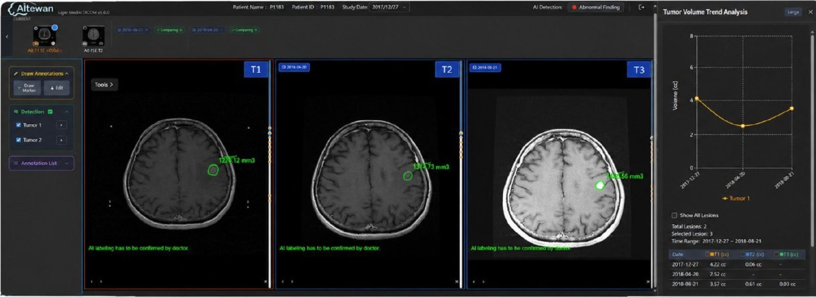
AI-assisted brain-tumor detection and initial contouring for radiotherapy planning. Compare prior and current MRI studies, review AI lesion marks, and track lesion volume over time.

FDA 510(k) K252190 TFDA No. 008460

DeepBT Detector-Plus viewer with three MRI timepoints and tumor volume trend
  1. 01Multi-timepoint MRICompare prior and current studies.
  2. 02AI lesion marksReview detected and contoured tumors.
  3. 03Volume trendTrack lesion volume over time.

TFDA clinical validation

  • 2 Taiwan medical centers
  • 133 cases · 181 lesions
  • Sensitivity 97.8% · Precision 88.9%
  • Dice 0.79 · Hausdorff 1.9% · Centroid distance 5.4%
  • Bi-parametric MRI · lesions ≥7 mm

FDA 510(k) standalone validation

  • 16 institutions (15 U.S. + 1 Taiwan)
  • 136 cases · 360 tumors
  • Sensitivity 88.6% · Dice 0.809
  • False positives 0.537 / case
  • Hausdorff 3.2% · Centroid 5.8%
  • Bi-parametric MRI · lesions ≥4 mm

Cleared in Taiwan and the U.S., with each performance claim tied to its own study design, lesion threshold and validation footprint.

IN CERTIFICATION

The next products in the portfolio.

Brain MRI

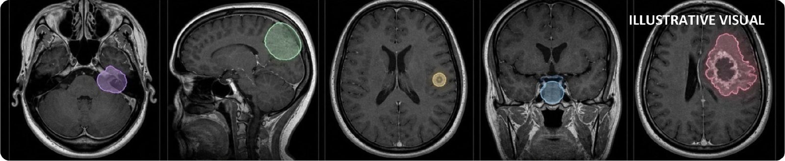
Five target brain-tumor types on MRI (illustrative)
  • Neuroma
  • Meningioma
  • Brain metastasis
  • Pituitary tumor
  • Glioblastoma

DeepBT® Detector A+

TFDA under review · not yet certified

Expanded detection, localization and five-class tumor classification (neuroma, meningioma, brain metastasis, pituitary tumor, glioblastoma) across multiple MRI input combinations.

Input: T1W+C alone or T1W+C + T2W. Lesions ≥0.4 cm.

96.30%
Lesion sensitivity
0.84
Dice
89.55%
5-class accuracy
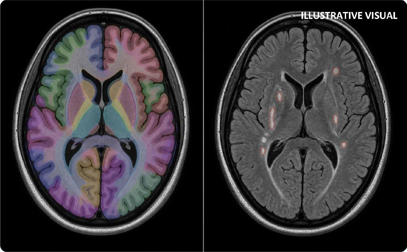
CereMap T1 structure map and FLAIR white-matter hyperintensities (illustrative)
  • T1 structures
  • FLAIR WMH
  • Dual models

CereMap

TFDA submission preparation · not yet certified

Sequence-specific analysis of gray-matter structures and white-matter hyperintensities. T1 and FLAIR are processed separately, one modality at a time, so the two are never mixed.

Output: DICOM-based segmentations and quantitative review data.

88.9%
T1 structure-wise Dice
78.0%
FLAIR WMH Dice
2
Sequence-specific models

Cardiovascular CT

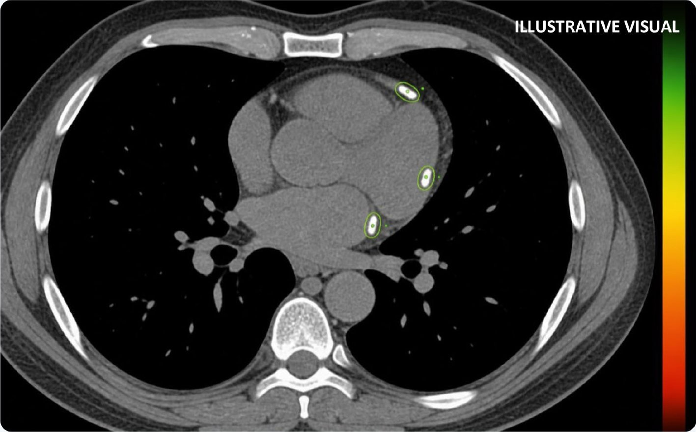
Coronary calcium foci on chest CT with risk scale (illustrative)
  • Calcium foci
  • Risk cue
  • Review

DeepLDCT-CAC

TFDA submission preparation · not yet certified

Automated coronary-artery calcium detection, Agatston scoring and risk stratification from chest CT, turning routine chest CT into an opportunity for structured cardiovascular risk discussion.

Input: low-dose or non-contrast chest CT (DICOM). Output: score, risk category, calcification area, imaging preview.

0.92
Binary sensitivity
0.93
Binary specificity
0–99 / ≥100
Low / high-risk grouping
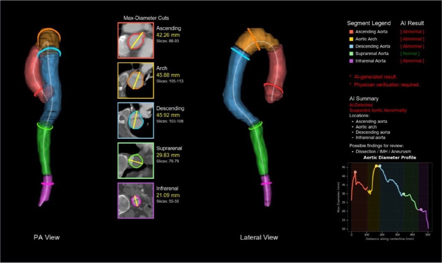
DeepAorA aortic segments, max-diameter cuts and diameter profile
  • Dissection
  • IMH
  • Aneurysm

DeepAorA Detector

TFDA under review · not yet certified

Case-level alerts for suspected aortic dissection, intramural hematoma and aortic aneurysm in adults ≥18 years on chest and abdominal CTA, with segmentation and assistive marks for physician review.

Input: complete-aorta CTA (DICOM). Output: case-level detection plus DICOM SC / SEG / PR / SR through Liger.

85.48%
Case-level sensitivity
88.60%
Case-level specificity
3
Target abnormality groups

Thoracic CT

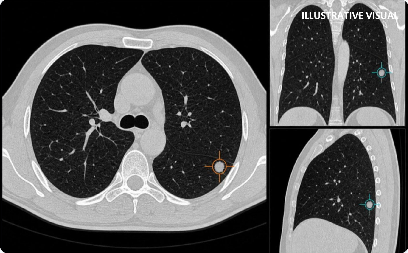
Lung nodule marked on axial, coronal and sagittal CT (illustrative)
  • Axial mark
  • Coronal view
  • Sagittal view

DeepPulmo

TFDA submission preparation · not yet certified

Two-stage lung-nodule and tumor detection (candidate generation, then 3D classification) designed for high sensitivity with false-positive reduction. Marks nodules 4–30 mm and masses >30 mm.

Input: axial chest CT or LDCT (DICOM), slice thickness ≤2.5 mm. Output: lesion marks, location, confidence, multiplanar review.

≈90%
Sensitivity in current materials
≈0.5
False positives / case
8,161
Development cases

Radiotherapy CT

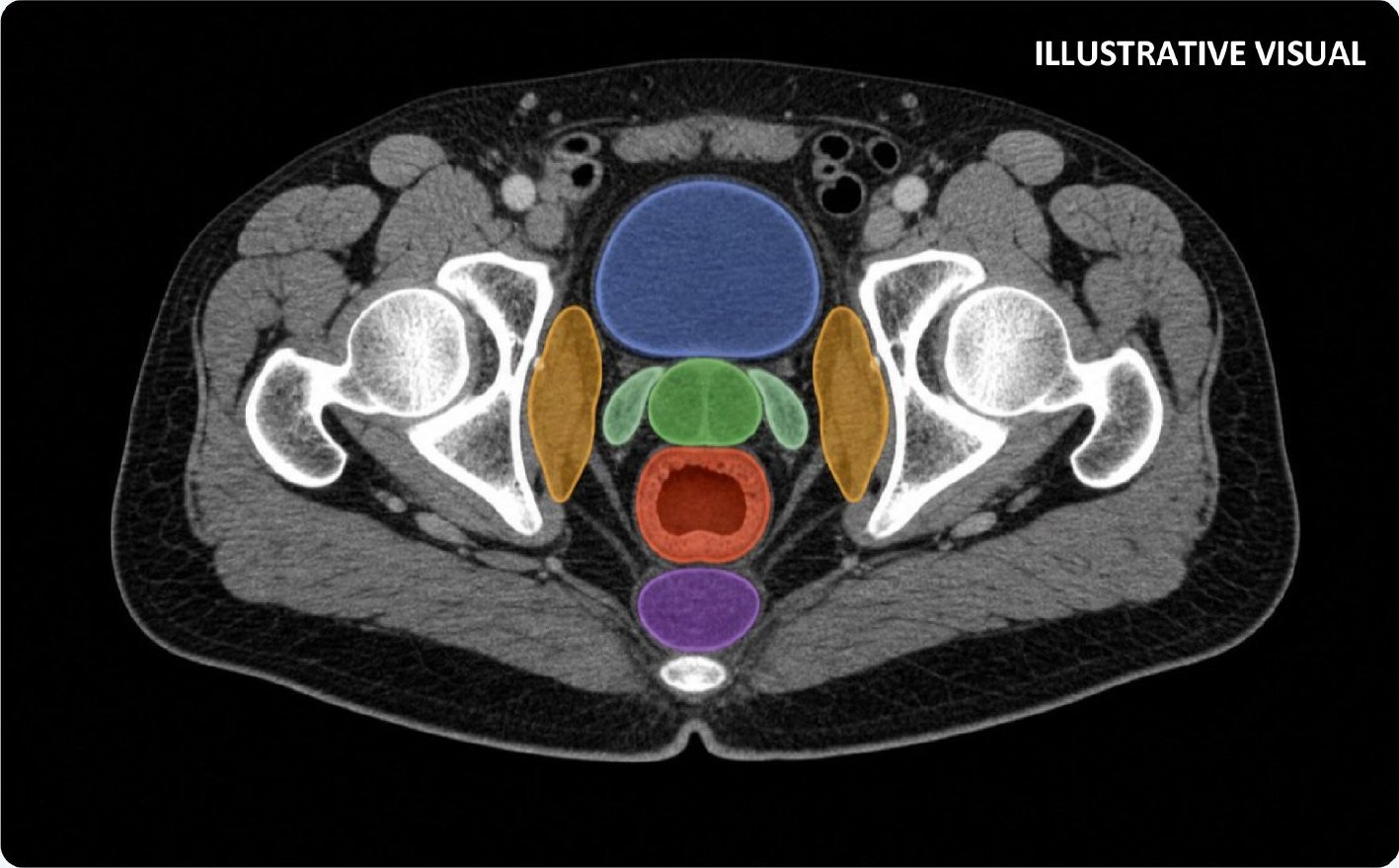
Pelvic CT with bladder, prostate/SV, rectum, nodes and OAR contours (illustrative)
  • Bladder
  • Prostate / SV
  • Rectum
  • Pelvic nodes
  • OAR

DeepPelvis

TFDA submission preparation · not yet certified

Automatic pelvic CTV and organ-at-risk segmentation (bladder, rectum, lymph nodes, prostate-related structures) giving a reviewable first contour set for radiotherapy planning.

Input: pelvic planning CT (DICOM). Supports PR / SEG / SC / SR and treatment-planning-system integration.

>0.75
CTV Dice target
>0.85
OAR Dice target
317
Development cohort cases

CLINICAL DEPLOYMENT

Designed for different hospital information environments.

One portfolio can meet hospitals where they are, without forcing every site into the same architecture.

01

Dedicated AI PACS

Imaging → AI router → Liger + AI PACS

Centralize AI results while leaving the hospital's core PACS unchanged.

02

Real-time PACS sync

Hospital PACS → Liger → PACS / viewer

Route studies in the background and return results to the normal reading workflow.

03

Upload + TPS

Planning CT → secure upload → Liger → TPS

Support radiotherapy planning through controlled transfer and TPS integration.

Regulatory status varies by market. Products marked under review or in preparation are not yet certified; images, specifications and content may be updated during development and regulatory review. AI outputs are assistive and support, not replace, clinical judgment.

Interested in Medical AI for your hospital or imaging network?

Tell us about your PACS environment and clinical domain, and we will connect you with the right product and deployment model.

Request Information →